REHOVOT, ISRAEL—October 27, 2020—Acute liver failure is a devastating, rapidly progressing disease that results in death in 80% of cases, unless an emergency liver transplant is performed. In the developed world, its leading cause is a substantial overdose of acetaminophen, also known as paracetamol.
In a study published in Nature Medicine, researchers from the labs of Prof. Eran Elinav and Prof. Ido Amit in the Weizmann Institute of Science’s Department of Immunology have, using mouse models of acute liver failure, discovered three liver-cell subsets that orchestrate the development of this condition. The scientists also identified signals – from the gut microbiome as well as the diseased liver – that jointly activate these cells, and showed that selectively blocking the signals and depleting the microbiome led to marked improvement in liver function and prolonged survival in the mice. An analysis of liver tissue from human patients with acute liver failure revealed a molecular pattern strikingly similar to the one identified in mice in the study, raising hopes that the findings may in the future be translated into a treatment for humans.
Dr. Aleksandra Kolodziejczyk, a postdoctoral fellow in Prof. Elinav’s lab, led the project in collaboration with other Weizmann Institute researchers and Dr. Amir Shlomai of the Liver Institute, Rabin Medical Center.
Dr. Kolodziejczyk and her colleagues began their investigation by creating gene expression profiles of 45,000 individual mouse liver cells, ultimately generating a comprehensive liver-cell atlas in conditions of health and acute failure. The scientists identified 49 cell subsets, of which three subsets of the stellate, endothelial and Kupffer cells became abnormally activated as the acute liver failure progressed in the mice. These previously undescribed cell subsets secreted a large variety of substances that attracted immune cells from outside the liver, which then contributed to its damage. All three subtypes shared a characteristic expression pattern of 77 genes – a pattern controlled by the same regulatory protein, the transcription factor MYC – thus suggesting that these cells may be activated through a common program.
The researchers suspected that the newly uncovered activation pathway could be regulated by signals from the gut microbiome. This makes anatomical sense, as the gastrointestinal tract drains into the liver through a large network of veins, directly exposing the liver to substances produced in the gut and by its microbes. When the scientists depleted the gut microbiome of the mice by administering wide-spectrum antibiotics, symptoms of liver failure were alleviated. Moreover, when the team induced acute liver failure in germ-free mice, who lack a microbiome, the condition was much less severe than in regular mice. Further studies of mice with and without a gut microbiome revealed that during acute liver failure, distinct molecules generated by the microbiome accumulate in the liver, where they activate the MYC protein in the three liver-cell subtypes that contribute to liver damage. In the absence of a microbiome, MYC activation was attenuated, leading to reduced liver damage.
Dr. Kolodziejczyk then worked out the molecular details of MYC activation, finding that the molecules coming from the microbiome activate the MYC program through surface receptors on the three liver-cell subtypes. She also found that the MYC program was activated in the same manner – that is, through the same receptors on the three cell subtypes – by signals coming from liver cells damaged by acetaminophen.
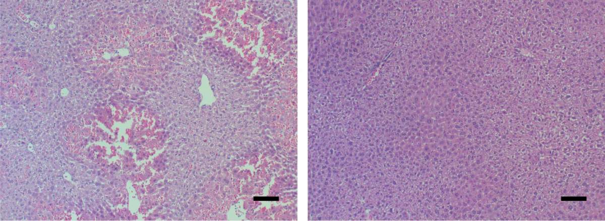
When the mice were genetically depleted of functioning receptors, given drugs that blocked MYC, or otherwise had the signals between the receptors and MYC interrupted, they no longer developed acute liver failure and their survival was extended. Gene expression analysis of individual cells showed that, in the treated mice, the three newly identified cell subtypes were no longer abnormally activated, and this reduced both the immune cell infiltration and the resultant liver damage.
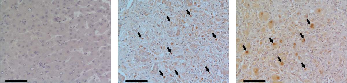
Finally, the researchers teamed up with Dr. Shlomai to analyze liver samples from patients with acute liver failure and compare them with samples from healthy liver donors. Samples from the patients – but not from healthy donors – were characterized by robust MYC activation similar to that observed in mice. These results raise the possibility that blocking the MYC program by drugs, coupled with microbiome modulation, may prove to be a potential treatment for acute liver failure.
“Our findings provide a first step towards achieving a comprehensive understanding of how the microbiome interacts with the host in contributing to acute liver failure,” Prof. Elinav says. “Such knowledge could lead to a new treatment option for this cureless and devastating disorder.”
Also taking part in the study were Dr. Sara Federici, Dr. Niv Zmora, Dr. Gayatree Mohapatra, Dr. Mally Dori-Bachash, Shanni Hornstein, Dr. Avner Leshem, and Dr. Hagit Shapiro of the Elinav lab; Dr. Tomer Meir Salame of Weizmann’s Life Sciences Core Facilities; Prof. Alon Harmelin of Weizmann’s Veterinary Resources Department; Dr. Debby Reuveni and Dr. Ehud Zigmond of the Tel Aviv Sourasky Medical Center; and Dr. Ana Tobar of the Rabin Medical Center.
Prof. Ido Amit’s research is supported by the Helen and Martin Kimmel Award for Innovative Investigation; the Sagol Institute for Longevity Research; the Kekst Family Institute for Medical Genetics; the Thompson Family Foundation Alzheimer’s Research Fund; the Adelis Foundation; Richard and Jacqui Scheinberg; the Ben B. and Joyce E. Eisenberg Foundation; the Anita James Rosen Foundation; the Lowy Foundation; the Wolfson Family Charitable Trust; the Vainboim Family; Lady Michelle Michels; Rosanne Cohen; Mauricio Gerson; Erika Mogyoros; Thomas Franklin Buchheim; Jeff Pinkner and Maya Iwanaga; the estate of Simon Saretzky; and the estate of Arthur Rath. Prof. Amit is the incumbent of the Eden and Steven Romick Professorial Chair.
Prof. Eran Elinav’s research is supported by the Morris Kahn Institute for Human Immunology; the Pearl Welinsky Merlo Scientific Progress Research Fund; the Hanna and Dr. Ludwik Wallach Cancer Research Fund; the Leona M. and Harry B. Helmsley Charitable Trust; the Adelis Foundation; the Else Kroener Fresenius Foundation; the Lawrence and Sandra Post Family Foundation; Yael and Rami Ungar; the Daniel Morris Trust; the Harold Altman Charitable Trust; the Howard and Nancy Marks Charitable Fund; the European Research Council; the Ben B. and Joyce E. Eisenberg Foundation; the Vainboim Family; Alex Davidoff; the White Rose International Foundation; and the Jeanne and Joseph Nissim Center for Life Sciences Research. Prof. Elinav is the incumbent of the Sir Marc and Lady Tania Feldmann Professorial Chair.

