
REHOVOT, ISRAEL—January 27, 2021—Chronic stress could be the prevailing condition of our time. In the short term, our jaws or stomachs may clench; in the long term, stress can lead to metabolic disease and speed up diseases of aging, as well as leading to more serious psychological disorders. The physical manifestations of stress originate in the brain, and they move along a so-called “stress axis” that ends in the adrenal glands. These glands then produce the hormone cortisol. When the stress axis is continually activated, changes occur in the cells and organs along the way, and the continual production of cortisol then substantially contributes to the symptoms of chronic stress.
The stress-response axis starts with the hypothalamus in the brain; moves through the pituitary gland, which is next to the brain; then advances to the adrenal glands, sited near the kidneys. Scientists at the Weizmann Institute of Science in Israel and the Max Planck Institute of Psychiatry in Germany have now used new technology to view the entire stress axis as it has never before been seen. Their findings, published in Science Advances, may be relevant to a number of stress-related diseases, ranging from anxiety and depression to metabolic syndrome and diabetes.
The new study, led by postdoctoral fellow Dr. Juan Pablo Lopez in Prof. Alon Chen’s joint neurobiology lab at the Weizmann Institute and Max Planck, made use of a relatively new technique that allows researchers to identify differences across all cell types in a tissue. This method is something like identifying the individual fruits in a bowl of fruit salad – rather than the standard method of turning that fruit salad into a “smoothie” and then trying to identify the average characteristics of all the fruits together. But in this case, the task was much more complex than simply separating apples from oranges: Dr. Pablo Lopez and the team mapped the entire length of the stress axis, checking the activities of numerous single cells all along the route. And they conducted this analysis on two sets of mice – one exposed to chronic stress, the other unstressed.
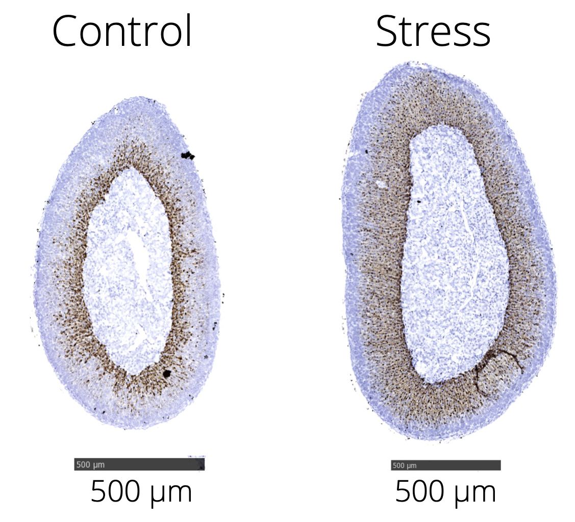
In total, the team mapped 21,723 cells along the three points in the stress axis, then compared their findings from the two sets of mice. They noted that as the stress message moved from one organ to the next, the gene expression in the cells and the tissues themselves underwent greater changes. The scientists identified 66 genes in the hypothalamus that were altered between normal and stressed mice, 692 in the pituitaries, and a whopping 922 in the adrenals. The adrenals are glands that can notably change size under chronic stress exposure, and it was here that the researchers noted the most significant alterations among the various cells.
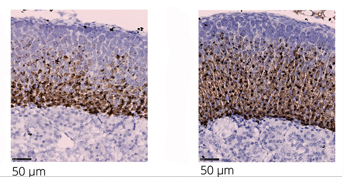
The unprecedented resolution of the technique enabled the team to identify, for the first time, a subpopulation of adrenal cells that may play a crucial role in stress response and adaptation. These were endocrine cells sitting in the outer layer, or adrenal cortex. Among other things, the scientists identified a gene, Abcb1b, that was overexpressed in these cells under stress situations. Abcb1b encodes a pump in the cell membrane that expels substances from the cell, and the scientists believe it plays a role in the release of cortisol. “If extra stress hormones are created, the cell needs extra release valves to let those hormones go,” says Dr. Pablo Lopez.
Are the findings in mice relevant to humans? In collaboration with researchers in university-based hospitals in the UK, Germany, Switzerland, and the US, the scientists obtained adrenal glands that had been removed from patients to relieve the symptoms of Cushing’s disease. Though the disease is the result of a growth on the pituitary, the result can be identical to chronic stress – weight gain and metabolic syndrome, high blood pressure, and depression or irritability – so in some cases it is treated by removing the adrenal glands, thereby reducing the patients’ stress hormone load. Indeed, the cells in these patients’ adrenals presented a picture similar to those of the mice in the chronic stress group.
The gene Abcb1 was known to the researchers from previous studies into the genetics of depression. It has been found that Abcb1 is polymorphic – meaning that it has several variants – and that at least one version is tied to a higher risk for depression. The group analyzed the expression of this variant in blood tests taken from a group of subjects who suffer from depression and who were subjected to temporary stress. They found that certain variants do indeed affect the ways the adrenal glands deal with stress signals coming down the axis.
Chronic stress can, of course, ultimately affect every part of the body and open the door to numerous health issues. Because it looks at the entire axis, on the one hand, and has mapped the axis down to the gene expression pattern of individual cells, on the other, the new study should provide a wealth of new information and insight into the mechanisms behind the stress axis. “Most research in this field has focused on chronic stress patterns in the brain,” says Prof. Chen. “In addition to presenting a possible new target for treating the diseases that arise from chronic stress, the findings of this study will open new directions for future research.”
Prof. Alon Chen’s research is supported by the Ruhman Family Laboratory for Research in the Neurobiology of Stress and by Bruno Licht. He is the incumbent of the Vera and John Schwartz Professorial Chair in Neurobiology.

