Improving Health & Medicine

Science Tips, September 2014

Scientists Identify the Signature of Aging in the Brain; Untangling the Maze; Waves of the Future


Scientists Identify the Signature of Aging in the Brain

Weizmann Institute researchers suggest that the brain’s “immunological age” is what counts

How the brain ages is still largely an open question – in part because this organ is mostly insulated from direct contact with other systems in the body, including the blood and immune systems. In research published recently in Science, Weizmann Institute researchers Prof. Michal Schwartz of the Department of Neurobiology and Dr. Ido Amit of the Department of Immunology found evidence of a unique “signature” that may be the “missing link” between cognitive decline and aging. The scientists believe that this discovery may, in the future, lead to treatments that can slow or reverse cognitive decline in older people.
Aging Brain Science Tips September

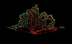
Immunofluorescence microscope image of the choroid plexus. Epithelial cells are in green and chemokine proteins (CXCL10) are in red

Until a decade ago, scientific dogma held that the blood-brain barrier prevents the blood-borne immune cells from attacking and destroying brain tissue. Yet in a long series of studies, Prof. Schwartz’s group showed that the immune system actually plays an important role both in healing the brain after injury and in maintaining the brain’s normal functioning. They have found that this brain-immune system interaction occurs across a barrier that is actually a unique interface within the brain’s territory.

This interface, known as the choroid plexus, is found in each of the brain’s four ventricles, and it separates the blood from the cerebrospinal fluid. Prof. Schwartz explains: “The choroid plexus acts as a ‘remote control’ for the immune system to affect brain activity. Biochemical ‘danger’ signals released from the brain are sensed through this interface; in turn, blood-borne immune cells assist by communicating with the choroid plexus. This cross-talk is important for preserving cognitive abilities and promoting the generation of new brain cells.” This finding led Prof. Schwartz and her group to suggest that cognitive decline over the years may be connected not only to one’s “chronological age,” but also to one’s “immunological age;” that is, changes in immune function over time might contribute to changes in brain function – not necessarily in step with the count of one’s years.

To test this theory, Prof. Schwartz and research students Kuti Baruch and Aleksandra Deczkowska teamed up with Dr. Amit and his research group. The researchers used next-generation sequencing technology to map changes in gene expression in 11 different organs, including the choroid plexus, in both young and aged mice, to identify and compare pathways involved in the aging process. That is how they identified a strikingly unique “signature of aging” that exists solely in the choroid plexus – not in the other organs. They discovered that one of the main elements of this signature was interferon beta – a protein that the body normally produces to fight viral infection. This protein appears to have a negative effect on the brain: When the researchers injected an antibody that blocks interferon beta activity into the cerebrospinal fluid of the older mice, their cognitive abilities were restored, as was their ability to form new brain cells. The scientists were also able to identify this unique signature in elderly human brains. The scientists hope that this finding may, in the future, help prevent or reverse cognitive decline in old age, by finding ways to rejuvenate the immunological age of the brain.

Dr. Ido Amit’s research is supported by the M.D. Moross Institute for Cancer Research; the J&R Center for Scientific Research; the Jeanne and Joseph Nissim Foundation for Life Sciences Research; the Abramson Family Center for Young Scientists; the Wolfson Family Charitable Trust; the Abisch Frenkel Foundation for the Promotion of Life Sciences; the Leona M. and Harry B. Helmsley Charitable Trust; Sam Revusky, Canada; the Florence Blau, Morris Blau and Rose Peterson Fund; the estate of Ernst and Anni Deutsch; the estate of Irwin Mandel; and the estate of David Levinson. Dr. Amit is the incumbent of the Alan and Laraine Fischer Career Development Chair.

Prof. Michal Schwartz’s research is supported by the Jeanne and Joseph Nissim Foundation for Life Sciences Research; the Adelis Foundation; the European Research Council; Nathan and Dora Oks, France; and Hilda Namm, Larkspur, CA. Prof. Schwartz is the incumbent of the Maurice and Ilse Katz Professorial Chair of Neuroimmunology.


Untangling the Maze

The fetus in the womb is wholly dependent on the blood bond with the mother. Spotting irregularities in the flow across the placenta could therefore be crucial for detecting fetal distress, but no reliable method is currently available for monitoring the flow or detecting other signs of the distress in its early stages.

Magnetic resonance imaging, or MRI, can be safely performed during pregnancy, but today’s MRI methods are not suitable, due to problems like the motion of the fetus or the mother’s breathing, the varied structure of placental tissue, and the tangled maze formed by maternal and fetal blood vessels.

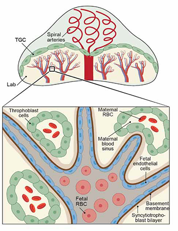
In a new study in mice conducted with advanced MRI methods, Weizmann Institute scientists have now revealed, in unprecedented detail, the dynamics of the flow of fluids within the placenta. This feat was all the more impressive given that a mouse placenta is around the size of a dime. As reported recently in the Proceedings of the National Academy of Sciences (PNAS), they managed to identify three different types of fluid-filled structures: maternal blood vessels, which account for two-thirds of the blood flow in the placenta; fetal vessels, which account for about one-quarter of the flow; and embryo-derived cells infiltrating the mother’s vasculature, which account for the rest of the flow and in which the exchange of fluids between mother and fetus takes place. The researchers also found that in maternal vessels, blood flows by diffusion, whereas in fetal vessels, the flow, stimulated by the pumping of the growing fetus’s heart, is much faster. In the cells that had infiltrated the mother’s vasculature, the dynamics of the flow follows an intermediate pattern, driven by both diffusion and pumping.

Two sophisticated MRI methods were combined to enable the study: one geared toward monitoring diffusion, and another directed at identifying structures with the help of a contrast material. They could be applied successfully in large part thanks to an innovative scanning approach, spatiotemporal encoding (SPEN), a Weizmann Institute technique. Because SPEN is ultrafast and makes it possible to separately encode signals from such different materials as air or fat, it allowed the researchers to overcome disturbances created by movement and the variability of placental tissue.
Click to enlarge photo.
Placenta Science Tips

Fluid-filled structures in the placenta: maternal and fetal blood vessels and embryo-derived trophoblast cells infiltrating the mother’s vasculature

If developed further for safe and reliable use in humans, this combined approach holds great promise as a noninvasive means of detecting fetal distress caused by disruptions in the placental flow. It can be particularly valuable when fast decisions about inducing labor need to be made; for example, with complications of pregnancy such as preeclampsia.

The study was a joint effort of two laboratories: one headed by Prof. Michal Neeman of the Department of Biological Regulation and the other by Prof. Lucio Frydman of the Department of Chemical Physics. The research was performed by two graduate students, Reut Avni from Prof. Neeman’s lab and Eddy Solomon from Prof. Frydman’s lab, together with Ron Hadas and Dr. Tal Raz of the Department of Biological Regulation and Dr. Peter Bendel of Chemical Research Support, in collaboration with Prof. Joel Richard Garbow from Washington University in St. Louis.

Prof. Lucio Frydman’s research is supported by the Helen and Martin Kimmel Institute for Magnetic Resonance Research, which he heads; the Helen and Martin Kimmel Award for Innovative Investigation; the Ilse Katz Institute for Material Sciences and Magnetic Resonance Research; the Adelis Foundation; the Mary Ralph Designated Philanthropic Fund of the Jewish Community Endowment Fund; Gary and Katy Leff, Calabasas, CA; Paul and Tina Gardner, Austin, TX; a Seventh Framework European Research Council Advanced Grant; and the Perlman Family Foundation.

Prof. Michal Neeman’s research is supported by the Henry Chanoch Krenter Institute for Biomedical Imaging and Genomics, which she heads; the Clore Center for Biological Physics, which she heads; the Kirk Center for Childhood Cancer and Immunological Disorders, which she heads; the Leona M. and Harry B. Helmsley Charitable Trust; the Carolito Stiftung; the Adelis Foundation; Andrew Adelson, Canada; the US-Israel Binational Science Foundation; and a Seventh Framework European Research Council Advanced Grant. Prof. Neeman is the incumbent of the Helen and Morris Mauerberger Professorial Chair in Biological Sciences.


Waves of the Future

In nature, waves – such as those in the ocean – begin as local oscillations in the water that spread out, ripple fashion, from their point of origin. But fans of Star Trek will recall a different sort of wave pattern: the tractor beam. Tractor beam technology, if it were to exist, would be based on waves that go in the opposite direction, converging from out in space onto the point of origin. In the show, the starship Enterprise would send out a tractor beam, like a cowboy’s lasso, to latch onto an object floating in space and pull it back toward the ship.

Prof. Gregory Falkovich of the Department of Physics of Complex Systems, working with the research group of Prof. Michael Shats of the National University of Australia, Canberra, recently showed that the idea may not be all science fiction.

In fact, the basic concept is rooted in the research of a 19th-century English physicist and mathematician, George Stokes, who examined the physics of waves in great detail. To do so, he placed small spheres in liquid and observed their movement in the fluid ripples. At the time, the prevailing theory held that if a wave was very small, a tiny sphere riding it would move within a closed circle. Stokes discovered that the sphere’s path would not be quite a closed circle. Instead, it would trace an inward-flowing spiral. This idea, which became known as “Stokes drift,” was assumed to be mostly theoretical. One might be able to create the conditions in a lab, but it would be hard, in nature, to find waves small enough to exhibit Stokes drift.

Prof. Falkovich’s collaborators, aided by observation technologies of the 21st century, returned to Stokes’s experiments to observe the particles that move in very small waves of light. They discovered, to their surprise, that a 3-D representation of the particles’ path is something like that of a drunken man making his way home. But, in their collaborative work, the researchers showed that not only is this movement not random, it is predictable – and can even be planned out ahead of time.

Using this insight, the scientists demonstrated the principle using two controlled fluid vortices. Between them, a wave flowed “backward” to the point at which the oscillations that created it originated. Some of the scientists in the group are already envisioning the creation of “tractor beams” in water that could, for example, latch onto pirate ships in the Indian Ocean and pull them in. More modest ideas (though still in the future) include using these waves to clean up pollution in the ocean.

Improving Health & Medicine

Science Tips, September 2014

• TAGS: Blood , Brain , Immune system , Physics , Technology

Scientists Identify the Signature of Aging in the Brain; Untangling the Maze; Waves of the Future


Scientists Identify the Signature of Aging in the Brain

Weizmann Institute researchers suggest that the brain’s “immunological age” is what counts

How the brain ages is still largely an open question – in part because this organ is mostly insulated from direct contact with other systems in the body, including the blood and immune systems. In research published recently in Science, Weizmann Institute researchers Prof. Michal Schwartz of the Department of Neurobiology and Dr. Ido Amit of the Department of Immunology found evidence of a unique “signature” that may be the “missing link” between cognitive decline and aging. The scientists believe that this discovery may, in the future, lead to treatments that can slow or reverse cognitive decline in older people.

Aging Brain Science Tips September

Immunofluorescence microscope image of the choroid plexus. Epithelial cells are in green and chemokine proteins (CXCL10) are in red

Until a decade ago, scientific dogma held that the blood-brain barrier prevents the blood-borne immune cells from attacking and destroying brain tissue. Yet in a long series of studies, Prof. Schwartz’s group showed that the immune system actually plays an important role both in healing the brain after injury and in maintaining the brain’s normal functioning. They have found that this brain-immune system interaction occurs across a barrier that is actually a unique interface within the brain’s territory.

This interface, known as the choroid plexus, is found in each of the brain’s four ventricles, and it separates the blood from the cerebrospinal fluid. Prof. Schwartz explains: “The choroid plexus acts as a ‘remote control’ for the immune system to affect brain activity. Biochemical ‘danger’ signals released from the brain are sensed through this interface; in turn, blood-borne immune cells assist by communicating with the choroid plexus. This cross-talk is important for preserving cognitive abilities and promoting the generation of new brain cells.” This finding led Prof. Schwartz and her group to suggest that cognitive decline over the years may be connected not only to one’s “chronological age,” but also to one’s “immunological age;” that is, changes in immune function over time might contribute to changes in brain function – not necessarily in step with the count of one’s years.

To test this theory, Prof. Schwartz and research students Kuti Baruch and Aleksandra Deczkowska teamed up with Dr. Amit and his research group. The researchers used next-generation sequencing technology to map changes in gene expression in 11 different organs, including the choroid plexus, in both young and aged mice, to identify and compare pathways involved in the aging process. That is how they identified a strikingly unique “signature of aging” that exists solely in the choroid plexus – not in the other organs. They discovered that one of the main elements of this signature was interferon beta – a protein that the body normally produces to fight viral infection. This protein appears to have a negative effect on the brain: When the researchers injected an antibody that blocks interferon beta activity into the cerebrospinal fluid of the older mice, their cognitive abilities were restored, as was their ability to form new brain cells. The scientists were also able to identify this unique signature in elderly human brains. The scientists hope that this finding may, in the future, help prevent or reverse cognitive decline in old age, by finding ways to rejuvenate the immunological age of the brain.

Dr. Ido Amit’s research is supported by the M.D. Moross Institute for Cancer Research; the J&R Center for Scientific Research; the Jeanne and Joseph Nissim Foundation for Life Sciences Research; the Abramson Family Center for Young Scientists; the Wolfson Family Charitable Trust; the Abisch Frenkel Foundation for the Promotion of Life Sciences; the Leona M. and Harry B. Helmsley Charitable Trust; Sam Revusky, Canada; the Florence Blau, Morris Blau and Rose Peterson Fund; the estate of Ernst and Anni Deutsch; the estate of Irwin Mandel; and the estate of David Levinson. Dr. Amit is the incumbent of the Alan and Laraine Fischer Career Development Chair.

Prof. Michal Schwartz’s research is supported by the Jeanne and Joseph Nissim Foundation for Life Sciences Research; the Adelis Foundation; the European Research Council; Nathan and Dora Oks, France; and Hilda Namm, Larkspur, CA. Prof. Schwartz is the incumbent of the Maurice and Ilse Katz Professorial Chair of Neuroimmunology.


Untangling the Maze

The fetus in the womb is wholly dependent on the blood bond with the mother. Spotting irregularities in the flow across the placenta could therefore be crucial for detecting fetal distress, but no reliable method is currently available for monitoring the flow or detecting other signs of the distress in its early stages.

Magnetic resonance imaging, or MRI, can be safely performed during pregnancy, but today’s MRI methods are not suitable, due to problems like the motion of the fetus or the mother’s breathing, the varied structure of placental tissue, and the tangled maze formed by maternal and fetal blood vessels.

In a new study in mice conducted with advanced MRI methods, Weizmann Institute scientists have now revealed, in unprecedented detail, the dynamics of the flow of fluids within the placenta. This feat was all the more impressive given that a mouse placenta is around the size of a dime. As reported recently in the Proceedings of the National Academy of Sciences (PNAS), they managed to identify three different types of fluid-filled structures: maternal blood vessels, which account for two-thirds of the blood flow in the placenta; fetal vessels, which account for about one-quarter of the flow; and embryo-derived cells infiltrating the mother’s vasculature, which account for the rest of the flow and in which the exchange of fluids between mother and fetus takes place. The researchers also found that in maternal vessels, blood flows by diffusion, whereas in fetal vessels, the flow, stimulated by the pumping of the growing fetus’s heart, is much faster. In the cells that had infiltrated the mother’s vasculature, the dynamics of the flow follows an intermediate pattern, driven by both diffusion and pumping.

Two sophisticated MRI methods were combined to enable the study: one geared toward monitoring diffusion, and another directed at identifying structures with the help of a contrast material. They could be applied successfully in large part thanks to an innovative scanning approach, spatiotemporal encoding (SPEN), a Weizmann Institute technique. Because SPEN is ultrafast and makes it possible to separately encode signals from such different materials as air or fat, it allowed the researchers to overcome disturbances created by movement and the variability of placental tissue.

Click to enlarge photo.
Placenta Science Tips

Fluid-filled structures in the placenta: maternal and fetal blood vessels and embryo-derived trophoblast cells infiltrating the mother’s vasculature

If developed further for safe and reliable use in humans, this combined approach holds great promise as a noninvasive means of detecting fetal distress caused by disruptions in the placental flow. It can be particularly valuable when fast decisions about inducing labor need to be made; for example, with complications of pregnancy such as preeclampsia.

The study was a joint effort of two laboratories: one headed by Prof. Michal Neeman of the Department of Biological Regulation and the other by Prof. Lucio Frydman of the Department of Chemical Physics. The research was performed by two graduate students, Reut Avni from Prof. Neeman’s lab and Eddy Solomon from Prof. Frydman’s lab, together with Ron Hadas and Dr. Tal Raz of the Department of Biological Regulation and Dr. Peter Bendel of Chemical Research Support, in collaboration with Prof. Joel Richard Garbow from Washington University in St. Louis.

Prof. Lucio Frydman’s research is supported by the Helen and Martin Kimmel Institute for Magnetic Resonance Research, which he heads; the Helen and Martin Kimmel Award for Innovative Investigation; the Ilse Katz Institute for Material Sciences and Magnetic Resonance Research; the Adelis Foundation; the Mary Ralph Designated Philanthropic Fund of the Jewish Community Endowment Fund; Gary and Katy Leff, Calabasas, CA; Paul and Tina Gardner, Austin, TX; a Seventh Framework European Research Council Advanced Grant; and the Perlman Family Foundation.

Prof. Michal Neeman’s research is supported by the Henry Chanoch Krenter Institute for Biomedical Imaging and Genomics, which she heads; the Clore Center for Biological Physics, which she heads; the Kirk Center for Childhood Cancer and Immunological Disorders, which she heads; the Leona M. and Harry B. Helmsley Charitable Trust; the Carolito Stiftung; the Adelis Foundation; Andrew Adelson, Canada; the US-Israel Binational Science Foundation; and a Seventh Framework European Research Council Advanced Grant. Prof. Neeman is the incumbent of the Helen and Morris Mauerberger Professorial Chair in Biological Sciences.


Waves of the Future

In nature, waves – such as those in the ocean – begin as local oscillations in the water that spread out, ripple fashion, from their point of origin. But fans of Star Trek will recall a different sort of wave pattern: the tractor beam. Tractor beam technology, if it were to exist, would be based on waves that go in the opposite direction, converging from out in space onto the point of origin. In the show, the starship Enterprise would send out a tractor beam, like a cowboy’s lasso, to latch onto an object floating in space and pull it back toward the ship.

Prof. Gregory Falkovich of the Department of Physics of Complex Systems, working with the research group of Prof. Michael Shats of the National University of Australia, Canberra, recently showed that the idea may not be all science fiction.

In fact, the basic concept is rooted in the research of a 19th-century English physicist and mathematician, George Stokes, who examined the physics of waves in great detail. To do so, he placed small spheres in liquid and observed their movement in the fluid ripples. At the time, the prevailing theory held that if a wave was very small, a tiny sphere riding it would move within a closed circle. Stokes discovered that the sphere’s path would not be quite a closed circle. Instead, it would trace an inward-flowing spiral. This idea, which became known as “Stokes drift,” was assumed to be mostly theoretical. One might be able to create the conditions in a lab, but it would be hard, in nature, to find waves small enough to exhibit Stokes drift.

Prof. Falkovich’s collaborators, aided by observation technologies of the 21st century, returned to Stokes’s experiments to observe the particles that move in very small waves of light. They discovered, to their surprise, that a 3-D representation of the particles’ path is something like that of a drunken man making his way home. But, in their collaborative work, the researchers showed that not only is this movement not random, it is predictable – and can even be planned out ahead of time.

Using this insight, the scientists demonstrated the principle using two controlled fluid vortices. Between them, a wave flowed “backward” to the point at which the oscillations that created it originated. Some of the scientists in the group are already envisioning the creation of “tractor beams” in water that could, for example, latch onto pirate ships in the Indian Ocean and pull them in. More modest ideas (though still in the future) include using these waves to clean up pollution in the ocean.