Improving Health & Medicine

No Cell Left Behind: Mapping the Human Body

Yale Scientific

Dots

Imagine walking into the doctor’s office, preparing for the worst. The doctor brings up surgery, vaguely motions to a chart on the wall, and points to certain printed organs. But what if he could show you which cells needed removal, what they looked like, and even why they caused your condition? Scientists may now have the means to determine the precise cellular structure of human organs, which could improve researchers’, doctors’, and patients’ understanding of human diseases.

Researchers at the Weizmann institute in Israel, led by Ido Amit and Shalev Itzkovitz, reconstructed the cellular structure of the liver in a Nature article published in February 2017. Their research team is a part of the Human Cell Atlas project, whose mission is to “create comprehensive reference maps of all human cells.” These maps, which organize cells by their genomes, will help researchers and medical professionals to better understand how and why cellular structures lead to organ functions.

While researchers have long been interested in cellular maps, the ability to determine the locations of cells and their genomic blueprints has been limited by available molecular biology techniques. Sequencing DNA, the alphabet of our genes, has traditionally been expensive and time-consuming. The cells must be extracted from the body before DNA can be harvested, destroying the spatial location of the cell.

Researchers also want to know the RNA profile of different cell types, which would reveal which genes are expressed as proteins. Ultimately, they would like to determine the proteome, or the entire set of expressed proteins, of each type of cell. However, this is still an emerging research field. Determining the levels of just 50 proteins in a cell is already a cumbersome task, let alone of the thousands of proteins actually present. The RNA profile serves as a simpler proxy for the proteome because modern advances in molecular biology, such as Next Generation Sequencing, have allowed researchers to quickly determine the DNA and RNA content of a cell. However, this process still requires the isolation of each cell prior to sequencing and sacrifices the cell’s spatial context in exchange for its genomic content.

The researchers at the Weizmann Institute were able to overcome this problem by combining the RNA sequencing results from Next Generation Sequencing with cellular locations determined by fluorescence. They then used computer algorithms to determine which RNA profile corresponded to which cellular location.

To do this, the researchers first determined the locations of several different cell types in the liver. One of the largest organs in the body, the liver is responsible for digesting nutrients and detoxifying dangerous substances. The cells in a subunit of the liver are differentiated into layers expanding radially from a central vein, and those closest to the vein are most accessible to the nutrients and oxygen carried in the bloodstream. The cells in each layer express a set of different genes, so the RNA profiles, which tell us which genes are expressed, differ between each layer.

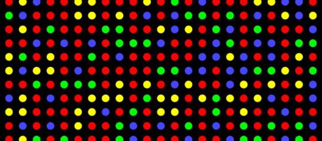
They then identified six “landmark” genes that are known to be expressed differently in each layer. They designed probes that would bind to each gene’s specific mRNA, a type of RNA that is translated into protein. With a technique called single molecule fluorescent in situ hybridization, the researchers determined which cells in each layer expressed which genes. “Each little dot corresponds to an mRNA,” said Thomas Pollard, a professor of Molecular, Cellular and Developmental Biology at Yale. A brighter dot signifies more copies of that gene’s mRNA. The Weizmann researchers now had data on six of the thousands of genes expressed in liver cells.

The next step was to find the entire RNA expression profile—the complete collection of different RNAs—for each layer. They turned to single cell-RNA sequencing to quickly identify the thousands of different RNAs within each cell. In this technique, RNA from each cell is harvested and amplified many times to allow it to be sequenced. Armed with this knowledge, the researchers compared the RNA expression levels of the six genes measured earlier. Since they knew the approximate expression levels of six of the genes from the previous experiment, they could match the RNA profiles to each cellular location from these six genes. For example, if landmark gene A was expressed highly in one RNA profile, then it would have to had come from a cell from a layer that fluoresced brightly for gene A’s mRNA.

Their maps showed that of the 7,277 genes expressed in liver cells, 3,496 of them vary non-randomly by spatial location. For example, genes in energy-demanding pathways were expressed the most near the central vein, which provides the cells with oxygen and nutrients. Cells near the vein also strongly expressed genes coding for secreted proteins; their placement near the vein allows cells to efficiently transport their secretory proteins through the vein. This finding confirmed the long-standing biological principle that structure leads to function.

Researchers would still like to dig further and determine the proteomes of different cells in addition to RNA profiles. Knowing which proteins are expressed at what levels in each cell would further illustrate the role of each cell in our body. “RNA expression profiles don’t give you protein levels,” Pollard explained. “Sometimes low mRNA levels can give you a lot of proteins, or a lot of mRNA can give you a few proteins. It also depends on the lifespan of the protein.”

Scott Holley, another professor of Molecular, Cellular and Developmental Biology at Yale, agrees. “It’s a caveat of the experiment,” Holley said. However, identifying the thousands of proteins in each cell requires thousands of antibodies to recognize and bind to each protein. This can be very difficult because researchers still do not know every protein our cells make. Finding every protein and producing antibodies for each one could take years.

Moreover, reference maps for other organs may not prove so easy. The cells in the liver are arranged in concentric circles, allowing the Weizmann researchers to assign cellular locations with as few as six genes. More complex structures, such as the brain, do not have such a simple geometry, making the reference map more complicated.

Nevertheless, cell maps will give researchers and medical professionals a new outlook on the human body. Embryo cell lineage mapping, a related technique used since the early 20th century, was a successful predecessor: it revealed the development of each cell in the embryos of various organisms. It has already provided important information about how organs and structures develop and given researchers the ability to manipulate embryonic organisms.

The Human Cell Atlas project hopes to continue to empower scientists and medical professionals, especially in cancer treatment. Rather than generalizing cancer to a whole organ, such as breast or lung cancer, this new blueprint may allow doctors and scientists to understand the inherent variation within each tumor. They can then more accurately monitor tumor growth and identify which therapy would be best suited for which cancers.

The lab at Weizmann is but one of many groups attempting to provide researchers with detailed maps of where each of the thousands of types of cells is located in the human body. The project is an international effort, with member laboratories in the United States, United Kingdom, Sweden, and Israel. Meanwhile, other mapping projects are also in the works. Cancer Research UK announced last month that a project to create an interactive virtual-reality map of breast cancers would receive up to $25 million. In the United States, the National Institute of Mental Health is preparing to announce grant awards for mapping mouse brains later this year.

There is still a long way to go to map out all 37 trillion cells in the human body, but with these advances in sequencing and imaging, it has never been easier to see our cells where they belong.

Improving Health & Medicine

No Cell Left Behind: Mapping the Human Body

Yale Scientific • TAGS: Proteins , Biology , Molecular genetics

Dots

Imagine walking into the doctor’s office, preparing for the worst. The doctor brings up surgery, vaguely motions to a chart on the wall, and points to certain printed organs. But what if he could show you which cells needed removal, what they looked like, and even why they caused your condition? Scientists may now have the means to determine the precise cellular structure of human organs, which could improve researchers’, doctors’, and patients’ understanding of human diseases.

Researchers at the Weizmann institute in Israel, led by Ido Amit and Shalev Itzkovitz, reconstructed the cellular structure of the liver in a Nature article published in February 2017. Their research team is a part of the Human Cell Atlas project, whose mission is to “create comprehensive reference maps of all human cells.” These maps, which organize cells by their genomes, will help researchers and medical professionals to better understand how and why cellular structures lead to organ functions.

While researchers have long been interested in cellular maps, the ability to determine the locations of cells and their genomic blueprints has been limited by available molecular biology techniques. Sequencing DNA, the alphabet of our genes, has traditionally been expensive and time-consuming. The cells must be extracted from the body before DNA can be harvested, destroying the spatial location of the cell.

Researchers also want to know the RNA profile of different cell types, which would reveal which genes are expressed as proteins. Ultimately, they would like to determine the proteome, or the entire set of expressed proteins, of each type of cell. However, this is still an emerging research field. Determining the levels of just 50 proteins in a cell is already a cumbersome task, let alone of the thousands of proteins actually present. The RNA profile serves as a simpler proxy for the proteome because modern advances in molecular biology, such as Next Generation Sequencing, have allowed researchers to quickly determine the DNA and RNA content of a cell. However, this process still requires the isolation of each cell prior to sequencing and sacrifices the cell’s spatial context in exchange for its genomic content.

The researchers at the Weizmann Institute were able to overcome this problem by combining the RNA sequencing results from Next Generation Sequencing with cellular locations determined by fluorescence. They then used computer algorithms to determine which RNA profile corresponded to which cellular location.

To do this, the researchers first determined the locations of several different cell types in the liver. One of the largest organs in the body, the liver is responsible for digesting nutrients and detoxifying dangerous substances. The cells in a subunit of the liver are differentiated into layers expanding radially from a central vein, and those closest to the vein are most accessible to the nutrients and oxygen carried in the bloodstream. The cells in each layer express a set of different genes, so the RNA profiles, which tell us which genes are expressed, differ between each layer.

They then identified six “landmark” genes that are known to be expressed differently in each layer. They designed probes that would bind to each gene’s specific mRNA, a type of RNA that is translated into protein. With a technique called single molecule fluorescent in situ hybridization, the researchers determined which cells in each layer expressed which genes. “Each little dot corresponds to an mRNA,” said Thomas Pollard, a professor of Molecular, Cellular and Developmental Biology at Yale. A brighter dot signifies more copies of that gene’s mRNA. The Weizmann researchers now had data on six of the thousands of genes expressed in liver cells.

The next step was to find the entire RNA expression profile—the complete collection of different RNAs—for each layer. They turned to single cell-RNA sequencing to quickly identify the thousands of different RNAs within each cell. In this technique, RNA from each cell is harvested and amplified many times to allow it to be sequenced. Armed with this knowledge, the researchers compared the RNA expression levels of the six genes measured earlier. Since they knew the approximate expression levels of six of the genes from the previous experiment, they could match the RNA profiles to each cellular location from these six genes. For example, if landmark gene A was expressed highly in one RNA profile, then it would have to had come from a cell from a layer that fluoresced brightly for gene A’s mRNA.

Their maps showed that of the 7,277 genes expressed in liver cells, 3,496 of them vary non-randomly by spatial location. For example, genes in energy-demanding pathways were expressed the most near the central vein, which provides the cells with oxygen and nutrients. Cells near the vein also strongly expressed genes coding for secreted proteins; their placement near the vein allows cells to efficiently transport their secretory proteins through the vein. This finding confirmed the long-standing biological principle that structure leads to function.

Researchers would still like to dig further and determine the proteomes of different cells in addition to RNA profiles. Knowing which proteins are expressed at what levels in each cell would further illustrate the role of each cell in our body. “RNA expression profiles don’t give you protein levels,” Pollard explained. “Sometimes low mRNA levels can give you a lot of proteins, or a lot of mRNA can give you a few proteins. It also depends on the lifespan of the protein.”

Scott Holley, another professor of Molecular, Cellular and Developmental Biology at Yale, agrees. “It’s a caveat of the experiment,” Holley said. However, identifying the thousands of proteins in each cell requires thousands of antibodies to recognize and bind to each protein. This can be very difficult because researchers still do not know every protein our cells make. Finding every protein and producing antibodies for each one could take years.

Moreover, reference maps for other organs may not prove so easy. The cells in the liver are arranged in concentric circles, allowing the Weizmann researchers to assign cellular locations with as few as six genes. More complex structures, such as the brain, do not have such a simple geometry, making the reference map more complicated.

Nevertheless, cell maps will give researchers and medical professionals a new outlook on the human body. Embryo cell lineage mapping, a related technique used since the early 20th century, was a successful predecessor: it revealed the development of each cell in the embryos of various organisms. It has already provided important information about how organs and structures develop and given researchers the ability to manipulate embryonic organisms.

The Human Cell Atlas project hopes to continue to empower scientists and medical professionals, especially in cancer treatment. Rather than generalizing cancer to a whole organ, such as breast or lung cancer, this new blueprint may allow doctors and scientists to understand the inherent variation within each tumor. They can then more accurately monitor tumor growth and identify which therapy would be best suited for which cancers.

The lab at Weizmann is but one of many groups attempting to provide researchers with detailed maps of where each of the thousands of types of cells is located in the human body. The project is an international effort, with member laboratories in the United States, United Kingdom, Sweden, and Israel. Meanwhile, other mapping projects are also in the works. Cancer Research UK announced last month that a project to create an interactive virtual-reality map of breast cancers would receive up to $25 million. In the United States, the National Institute of Mental Health is preparing to announce grant awards for mapping mouse brains later this year.

There is still a long way to go to map out all 37 trillion cells in the human body, but with these advances in sequencing and imaging, it has never been easier to see our cells where they belong.